Publications
The Psychology of Burnout: Stress, Sleep and Cognition
Abstract
Burnout is usually described in psychological terms, but patients describe it in their bodies: they cannot sleep, cannot concentrate, cannot remember, and a night's rest no longer restores them. This article reads the research available in January 2022 from a psychologist's point of view: how the appraisal of stress, rumination and the failure to recover turn chronic work pressure into a state that can be measured in hormones, sleep, thinking and the brain. Despite decades of work on the hypothalamic–pituitary–adrenal (HPA) axis, no reliable cortisol marker of burnout has been found. Sleep findings are more consistent: short and fragmented sleep both predicts and accompanies clinical burnout. Cognitive deficits are well documented. A 2021 meta-analysis of 17 studies found small to moderate impairments in episodic memory, working memory, executive function, attention and processing speed in patients with clinical burnout compared with healthy controls. Brain imaging studies from Stockholm report thinning of the medial prefrontal cortex and enlarged amygdalae in people with chronic occupational stress. Much of this evidence comes from Sweden, where exhaustion disorder (utmattningssyndrom, ICD-10-SE F43.8A) has been an official diagnosis since 2005 and has become a leading cause of sick leave. Follow-up studies show that recovery is slow: seven years after seeking care, a third of patients are still clinically exhausted.
Keywords: burnout; stress appraisal; rumination; recovery; exhaustion disorder; HPA axis; cortisol; sleep; cognition; executive function; Sweden; sick leave
Introduction: from Selye to allostatic load
The biology of stress has a long history. In 1915 Walter Cannon described the "fight or flight" response, the rapid activation of the sympathetic nervous system and the release of adrenaline (Cannon, 1915). In 1936 Hans Selye, then a young researcher in Montreal, reported in Nature that rats exposed to very different harmful agents (cold, surgical injury, toxic substances) developed the same pattern: enlarged adrenal glands, shrinking of the thymus and lymph nodes, and stomach ulcers (Selye, 1936). He called it the general adaptation syndrome and described three phases: alarm, resistance and, if the stressor persisted, exhaustion.
Selye's third phase is the closest early biological description of what we now call burnout. His idea that the stress response is general and non-specific has since been heavily revised. In 1998 Bruce McEwen proposed the concept of allostatic load: the cumulative cost to the body of repeated or prolonged activation of the systems that help it adapt (McEwen, 1998). The stress response is protective in the short term. Kept switched on for months, or unable to switch off, it starts to cause harm.
Psychology adds the missing link between the stressor and the body. In Lazarus and Folkman's (1984) transactional model, a situation becomes stressful when a person appraises it as a threat and judges their resources insufficient to cope. The body responds to that appraisal rather than to the workload as such. Brosschot, Gerin and Thayer (2006) added a second step: perseverative cognition. Worry and rumination keep the stress response active long after the stressor has gone, in the evening, at the weekend, at three in the morning. Burnout, seen this way, is less the result of hard work than of hard work that the mind cannot leave behind.
With this frame in mind, the rest of the article follows the path from mind to body: the hormonal stress axis, sleep, thinking, the brain circuits of emotion regulation, and the long road to recovery.
The HPA axis: the search for a cortisol marker
The hypothalamic–pituitary–adrenal axis is the body's slow stress system. When the brain registers a demand, the hypothalamus releases corticotropin-releasing hormone (CRH), the pituitary responds with adrenocorticotropic hormone (ACTH), and the adrenal cortex releases cortisol. Cortisol mobilises energy, modulates the immune system, and feeds back on the brain to switch the response off.
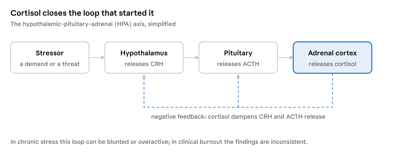
It seemed obvious that burnout, a state of exhaustion after prolonged stress, would leave a trace in this system. Researchers have measured cortisol in saliva, blood, urine and hair; they have looked at the morning rise after waking (the cortisol awakening response), the slope across the day, and the response to laboratory stress tests.
The results have been disappointing. A systematic review found no biomarker of burnout, largely because studies were not comparable: they defined burnout in different ways, measured biomarkers differently and controlled for different confounders (Danhof-Pont, van Veen & Zitman, 2011). A later narrative review by the Gothenburg Institute of Stress Medicine reached the same conclusion: research cannot confirm any consistent endocrine or immune change in burnout, although acute reactivity of the HPA axis and of anabolic hormones may be altered in clinical burnout (Jonsdottir & Sjörs Dahlman, 2019). Studies of thyroid hormones, prolactin and growth hormone were inconclusive.
There are good reasons for this. Cortisol varies widely across the day and between people, and depends on sleep, medication, smoking, menstrual cycle and the exact time of sampling. Many studies used questionnaire scores in working samples rather than clinical cases. And the HPA axis can be overactive in one phase of chronic stress and blunted in another, so groups at different stages may cancel each other out. In practice, there is no laboratory test for burnout, and a normal cortisol result neither confirms nor excludes it.
Sleep: cause and consequence
Where cortisol has disappointed, sleep has not. Patients with clinical burnout sleep badly, and the disturbance can be measured. In polysomnographic recordings, Ekstedt and colleagues (2006) found more arousals and fragmented sleep, more time awake and in light stage-1 sleep, lower sleep efficiency, and less slow-wave and REM sleep in burnout patients than in controls. Slow-wave sleep is the deepest and most restorative stage, so its loss fits the patients' complaint that sleep no longer refreshes them.
Sleep problems also come before burnout. In a prospective study at a Swedish workplace, 15 of 388 employees developed clinical burnout over two years. Sleeping less than six hours a night was the main baseline predictor, after adjustment for work demands, thoughts of work during leisure time and sleep quality (Söderström, Jeding, Ekstedt, Perski & Åkerstedt, 2012). The authors suggested a chain: high demands lead to work-related thoughts in the evening, which shorten and disturb sleep, which in turn prevents recovery. The middle link is psychological. Research on recovery shows that time off protects people when it comes with psychological detachment, the experience of mentally switching off from work during leisure time (Sonnentag & Fritz, 2015). Rumination is its opposite, and it is one of the most treatable parts of the chain.
Recovery follows the same path in reverse. In follow-up recordings six to twelve months later, patients whose burnout improved also showed better sleep quality and daytime alertness (Ekstedt, Söderström & Åkerstedt, 2009). Grossi et al. (2015), reviewing the clinical literature, concluded that sleep impairments are both causative and maintaining factors in clinical burnout. This makes sleep one of the few biological targets that can be changed directly.
Cognition: what burnout does to thinking
Ask patients with clinical burnout what troubles them most and many will not mention tiredness first. They describe losing the thread of a conversation, reading a page three times, forgetting appointments, being unable to plan a simple task. Until recently it was not clear how much of this showed up on neuropsychological tests.
A meta-analysis published in December 2021 settled much of the question (Gavelin et al., 2022). It pooled 17 studies with 730 patients with clinical burnout and 649 healthy controls (Figure 2).
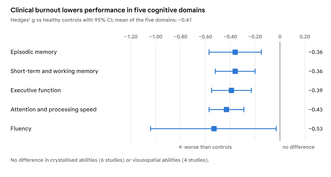
The deficits are small to moderate, around 0.4 standard deviations, and they fall on functions that depend on effortful control: holding information in mind, switching between tasks, keeping attention on a target, retrieving recent events. Knowledge and skills learned long ago (crystallised abilities) and visuospatial abilities are intact. This profile fits the idea that burnout drains the capacity for sustained mental effort rather than damaging stored knowledge.
Two cautions. First, 0.4 standard deviations is a group average: some patients perform normally, others are clearly impaired. Second, test scores in a quiet room capture only part of the problem. Patients often say that the real difficulty is mental fatigue that sets in after a short period of effort, which a 20-minute test does not measure.
The deficits also last. In a Gothenburg cohort, working memory and attention were still impaired three years after patients first sought care (Jonsdottir et al., 2017). Seven to twelve years later, former patients who no longer met the diagnostic criteria still reported more cognitive problems and fatigue than healthy controls (Ellbin, Jonsdottir, Eckerström & Eckerström, 2021).
The brain: what imaging studies show
Most imaging work on burnout comes from one research group at the Karolinska Institutet in Stockholm, led by Ivanka Savic, so the findings need replication. They are nonetheless consistent with each other and with animal research on chronic stress (Table 1).
| Study | Participants | Main finding |
|---|---|---|
| Blix, Perski, Berglund & Savic (2013), PLOS ONE | People with long-term occupational stress vs controls | Reduced grey matter volume in the anterior cingulate cortex and dorsolateral prefrontal cortex |
| Savic (2015), Cerebral Cortex | People with chronic occupational stress vs controls | Thinning of the medial prefrontal cortex; the normal age-related thinning of the frontal cortex was more pronounced; larger amygdalae, with volume correlating with perceived stress |
| Golkar et al. (2014), PLOS ONE | People with work-related chronic stress vs controls | Weaker functional coupling between the amygdala and the medial prefrontal cortex; more difficulty down-regulating negative emotion |
Table 1. Brain imaging studies of chronic occupational stress from the Karolinska Institutet.
The pattern makes sense. The medial prefrontal cortex helps regulate emotional responses generated in the amygdala. If the regulator is thinner and less connected, and the alarm system is larger, stressful events would be harder to switch off. That is close to what patients describe: they react more strongly to small irritations and calm down more slowly.
Whether these changes cause burnout, result from it, or both, cannot be answered by cross-sectional studies. Savic's group reported that some of the changes were partially reversed after treatment and recovery (Savic, Perski & Osika, 2018), which, if confirmed, would mean the brain changes are a state rather than a permanent scar.
Sweden's exhaustion disorder: burnout as a diagnosis
In the late 1990s Sweden saw a sharp rise in long-term sick leave for stress-related problems. Doctors had no adequate diagnosis: burnout was not a medical term, and adjustment disorder did not capture the central role of fatigue. In 2003 an expert group convened by the National Board of Health and Welfare (Socialstyrelsen) proposed criteria for a new diagnosis, utmattningssyndrom, usually translated as exhaustion disorder. In 2005 it entered the Swedish version of ICD-10 as code F43.8A, a specification of "other reactions to severe stress" (Socialstyrelsen, 2003). It exists only in Sweden.
| Criterion | Content (abridged) |
|---|---|
| A | Physical and mental symptoms of exhaustion for at least two weeks, developed in response to one or more identifiable stressors present for at least six months |
| B | Markedly reduced mental energy: lack of initiative, lack of endurance, or a longer recovery time after mental effort |
| C | At least four of the following, nearly every day for the same two weeks: concentration or memory problems; markedly reduced ability to cope with demands or time pressure; emotional instability or irritability; sleep disturbance; marked physical weakness or fatigability; physical symptoms such as pain, palpitations, gastrointestinal problems, dizziness or sound sensitivity |
| D | Clinically significant distress or impaired functioning at work, socially or in other areas |
| E | Not due to a substance or a general medical condition |
| F | If criteria for major depression, dysthymia or generalised anxiety are met, exhaustion disorder is coded as a comorbid condition |
Table 2. Diagnostic criteria for exhaustion disorder (ICD-10-SE F43.8A), abridged. Source: Socialstyrelsen (2003).
Two features distinguish exhaustion disorder from the psychological concept of burnout. It is not restricted to work: the stressors can be private. And it puts cognitive symptoms and the loss of mental energy at the centre, rather than cynicism or reduced professional efficacy. Grossi et al. (2015) concluded that it is the most valid clinical equivalent of burnout currently available.
The diagnosis spread fast. Between 2010 and 2015 the number of new sick-leave cases in Sweden rose by 98,000; psychiatric diagnoses accounted for 57,000 of them, or 59% of the increase, and stress reactions grew the most (Försäkringskassan, 2016). By the mid-2010s, adjustment disorders and reactions to severe stress, the group that includes exhaustion disorder, accounted for about half of all new sick-leave cases with a psychiatric diagnosis (Försäkringskassan, 2017). In the municipal and regional sector, 37.4% of new long sick-leave cases (over 90 days) in 2017–2018 were due to mental diagnoses, and four out of five of these were stress reactions or depression (AFA Försäkring, n.d.).
The diagnosis has critics. Its criteria overlap with depression, anxiety and chronic pain, and it has been adopted in no other country. ICD-11, in force internationally since 1 January 2022, has no equivalent category, which raises the question of what will happen to it when Sweden moves to the new classification.
Recovery: slow and often incomplete
The Swedish cohorts have one great advantage: they follow patients for years. The results are sobering (Table 3).
| Follow-up | Finding | Source |
|---|---|---|
| 3 years | Working memory and attention still impaired | Jonsdottir et al. (2017) |
| 7 years (217 former patients, 74% women) | 46% still report extreme fatigue; 73% report reduced stress tolerance; on clinical assessment, one in three is still judged to have exhaustion | Glise, Wiegner & Jonsdottir (2020) |
| 7–12 years | Patients who still meet the criteria report wide-ranging cognitive and daily-life problems; even those considered recovered report more cognitive problems and fatigue than healthy controls | Ellbin et al. (2021) |
Table 3. Long-term follow-up of patients with exhaustion disorder in Sweden.
Men and women showed similar patterns of residual symptoms (Glise et al., 2020). The main lasting complaint is reduced tolerance to stress, more than low mood: the person functions, but has a narrower margin before symptoms return. Many patients describe changing their working life to fit it. In a qualitative follow-up ten years after diagnosis, former patients described recovery as a long process that also brought a more authentic self-image, alongside lasting cognitive difficulties and reduced energy (Ellbin, Jonsdottir & Bååthe, 2021).
Implications for practice
Burnout is a psychological process with a biological footprint. It is less mysterious than it looked twenty years ago and less spectacular than the popular image of "cortisol burnout". There is no single biomarker. There is a well-documented pattern of disturbed sleep, reduced cognitive capacity and changes in the brain circuits that regulate stress, with a recovery that takes years rather than weeks. The point of entry for treatment remains psychological: how the person appraises demands, whether they can stop thinking about work, and whether they can rebuild a rhythm of effort and rest.
I see clients in English, French and Italian, at the practice in Gasperich (Luxembourg) on Saturdays or by video consultation on weekday evenings.
Request an appointmentReferences
- AFA Försäkring. (n.d.). Psykisk ohälsa i län: Långvarig sjukfrånvaro med psykisk diagnos 2017–2018 (F6389). https://www.afaforsakring.se/dokument/tytshmx8vzhbwhzdizsp/f6389_psykisk-ohalsa-lan.pdf
- Blix, E., Perski, A., Berglund, H., & Savic, I. (2013). Long-term occupational stress is associated with regional reductions in brain tissue volumes. PLOS ONE, 8(6), e64065. https://doi.org/10.1371/journal.pone.0064065
- Brosschot, J. F., Gerin, W., & Thayer, J. F. (2006). The perseverative cognition hypothesis: A review of worry, prolonged stress-related physiological activation, and health. Journal of Psychosomatic Research, 60(2), 113–124. https://doi.org/10.1016/j.jpsychores.2005.06.074
- Cannon, W. B. (1915). Bodily changes in pain, hunger, fear and rage. D. Appleton.
- Danhof-Pont, M. B., van Veen, T., & Zitman, F. G. (2011). Biomarkers in burnout: A systematic review. Journal of Psychosomatic Research, 70(6), 505–524. https://doi.org/10.1016/j.jpsychores.2010.10.012
- Ekstedt, M., Söderström, M., Åkerstedt, T., Nilsson, J., Søndergaard, H.-P., & Perski, A. (2006). Disturbed sleep and fatigue in occupational burnout. Scandinavian Journal of Work, Environment & Health, 32(2), 121–131. https://doi.org/10.5271/sjweh.987
- Ekstedt, M., Söderström, M., & Åkerstedt, T. (2009). Sleep physiology in recovery from burnout. Biological Psychology, 82(3), 267–273. https://doi.org/10.1016/j.biopsycho.2009.08.006
- Ellbin, S., Jonsdottir, I. H., & Bååthe, F. (2021). "Who I am now, is more me." An interview study of patients' reflections 10 years after exhaustion disorder. Frontiers in Psychology, 12, 752707. https://doi.org/10.3389/fpsyg.2021.752707
- Ellbin, S., Jonsdottir, I. H., Eckerström, C., & Eckerström, M. (2021). Self-reported cognitive impairment and daily life functioning 7–12 years after seeking care for stress-related exhaustion. Scandinavian Journal of Psychology, 62(4), 484–493. https://doi.org/10.1111/sjop.12722
- Försäkringskassan. (2016). Sjukskrivning för reaktioner på svår stress ökar mest (Korta analyser 2016:2). Försäkringskassan.
- Försäkringskassan. (2017). Psykiatriska diagnoser (Korta analyser 2017:1). Försäkringskassan.
- Gavelin, H. M., Domellöf, M. E., Åström, E., Nelson, A., Launder, N. H., Stigsdotter Neely, A., & Lampit, A. (2022). Cognitive function in clinical burnout: A systematic review and meta-analysis. Work & Stress, 36(1), 86–104. https://doi.org/10.1080/02678373.2021.2002972 (published online 3 December 2021)
- Glise, K., Wiegner, L., & Jonsdottir, I. H. (2020). Long-term follow-up of residual symptoms in patients treated for stress-related exhaustion. BMC Psychology, 8, 26. https://doi.org/10.1186/s40359-020-0395-8
- Golkar, A., Johansson, E., Kasahara, M., Osika, W., Perski, A., & Savic, I. (2014). The influence of work-related chronic stress on the regulation of emotion and on functional connectivity in the brain. PLOS ONE, 9(9), e104550. https://doi.org/10.1371/journal.pone.0104550
- Grossi, G., Perski, A., Osika, W., & Savic, I. (2015). Stress-related exhaustion disorder – clinical manifestation of burnout? A review of assessment methods, sleep impairments, cognitive disturbances, and neuro-biological and physiological changes in clinical burnout. Scandinavian Journal of Psychology, 56(6), 626–636. https://doi.org/10.1111/sjop.12251
- Jonsdottir, I. H., Nordlund, A., Ellbin, S., Ljung, T., Glise, K., Währborg, P., Sjörs, A., & Wallin, A. (2017). Working memory and attention are still impaired after three years in patients with stress-related exhaustion. Scandinavian Journal of Psychology, 58(6), 504–509. https://doi.org/10.1111/sjop.12394
- Jonsdottir, I. H., & Sjörs Dahlman, A. (2019). Mechanisms in endocrinology: Endocrine and immunological aspects of burnout: A narrative review. European Journal of Endocrinology, 180(3), R147–R158. https://doi.org/10.1530/EJE-18-0741
- Lazarus, R. S., & Folkman, S. (1984). Stress, appraisal, and coping. Springer.
- McEwen, B. S. (1998). Protective and damaging effects of stress mediators. New England Journal of Medicine, 338(3), 171–179. https://doi.org/10.1056/NEJM199801153380307
- Savic, I. (2015). Structural changes of the brain in relation to occupational stress. Cerebral Cortex, 25(6), 1554–1564. https://doi.org/10.1093/cercor/bht348
- Savic, I., Perski, A., & Osika, W. (2018). MRI shows that exhaustion syndrome due to chronic occupational stress is associated with partially reversible cerebral changes. Cerebral Cortex, 28(3), 894–906. https://doi.org/10.1093/cercor/bhw413
- Selye, H. (1936). A syndrome produced by diverse nocuous agents. Nature, 138, 32. https://doi.org/10.1038/138032a0
- Socialstyrelsen. (2003). Utmattningssyndrom: Stressrelaterad psykisk ohälsa. Bokförlaget Bilda.
- Sonnentag, S., & Fritz, C. (2015). Recovery from job stress: The stressor-detachment model as an integrative framework. Journal of Organizational Behavior, 36(S1), S72–S103. https://doi.org/10.1002/job.1924
- Söderström, M., Jeding, K., Ekstedt, M., Perski, A., & Åkerstedt, T. (2012). Insufficient sleep predicts clinical burnout. Journal of Occupational Health Psychology, 17(2), 175–183. https://doi.org/10.1037/a0027518
© 2022 Giampaolo Penzo. All rights reserved. No reproduction, translation or reuse, including for AI training, without the author's written permission. Short quotations are welcome with credit and a link to this page. Legal notice