Publications
The Anxious Brain: Neurobiology of Fear and Worry
Key points
- Fear and anxiety use overlapping but distinct circuits. The central amygdala drives fast responses to imminent threat; the bed nucleus of the stria terminalis (BNST) sustains vigilance when the threat is uncertain.
- The ventromedial prefrontal cortex acts as a brake on the amygdala. It is central to fear extinction, the process that exposure therapy relies on.
- Neuroimaging meta-analyses show amygdala and insula hyperactivity across several anxiety disorders, but no scan can yet diagnose one.
- Twin studies put the heritability of panic disorder at about 43% and of generalised anxiety disorder at about 32%. The rest comes mostly from experiences that family members do not share.
- Early adversity can leave chemical marks on stress-related genes. This is one route by which experience gets under the skin.
Introduction
People with panic disorder often say their body reacts before they have decided anything is wrong. Neuroscience agrees with them. Much of the defensive response starts in structures that work faster than conscious thought and that we share with other mammals. Conscious worry, the part people usually call anxiety, sits on top of that machinery and talks back to it.
This article follows the threat signal through the brain: from the amygdala, which detects it, to the prefrontal cortex, which can turn it down, to the hormones and neurotransmitters that carry it and finally to the genes and early experiences that set how sensitive the whole system is. A warning before starting: most circuit-level knowledge comes from rodents. Human imaging confirms the broad picture, but the details are less certain than textbook diagrams suggest.
1. Fear and anxiety in the brain
The amygdala is often called the brain's fear centre. The label is half right. The amygdala is a cluster of nuclei with different jobs and it sits inside a wider network that also handles anxiety.
Threat information reaches the basolateral amygdala (BLA) from the thalamus and the sensory cortices. The BLA is where a neutral cue, such as a tone or a face, becomes associated with danger. The hippocampus adds context: the same dog is a threat in a dark alley and harmless in a friend's living room. From the BLA the signal goes to two output stations. The central amygdala (CeA) triggers rapid, short-lived defence: freezing, a jump in heart rate, a startle. The bed nucleus of the stria terminalis (BNST), part of what anatomists call the extended amygdala, produces slower and longer-lasting states of vigilance (Davis et al., 2010; Tovote et al., 2015).
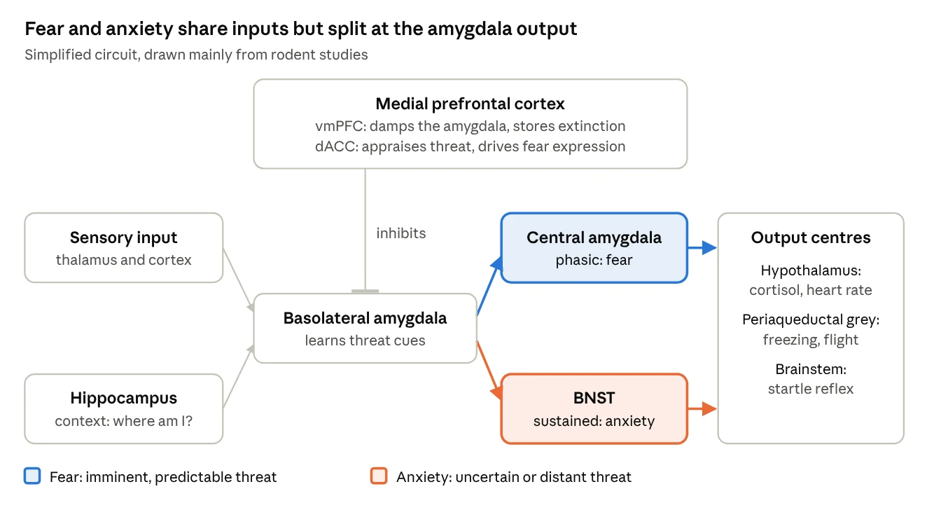
The split matches the behavioural distinction between fear and anxiety. In rodents, lesions of the CeA abolish responses to a cue that predicts a shock within seconds, while lesions of the BNST reduce the sustained responses seen when the threat is diffuse or unpredictable (Davis et al., 2010). Human imaging is harder, because the BNST is tiny, but a meta-analysis of human studies found that the BNST is engaged during the anticipation of uncertain threat (Avery et al., 2016).
The clean division in the figure is a simplification. Reviewing human and monkey studies, Shackman and Fox (2016) point out that the central amygdala and the BNST are tightly interconnected, that both respond to brief and to sustained threat and that both can drive fear and anxiety. They treat the two as one functional unit, the central extended amygdala, rather than as separate fear and anxiety modules (Shackman & Fox, 2016). The question is still open.
Uncertainty is the key variable. One influential model proposes that what anxious people share is an exaggerated anticipatory response when the likelihood, timing or nature of a threat is unknown (Grupe & Nitschke, 2013). This fits clinical experience. People with generalised anxiety disorder rarely fear one specific thing. What they cannot tolerate is not knowing.
One caution on terms. LeDoux and Pine (2016) argue that these circuits control defensive behaviour and bodily responses, while the conscious feeling of fear or anxiety depends on cortical systems that are only partly understood (LeDoux & Pine, 2016). The rat that freezes and the person who feels afraid may share the circuit for the first part but not necessarily for the second.
2. The prefrontal brake
The amygdala does not work alone. The medial prefrontal cortex and the anterior cingulate, on the inner surface of the frontal lobes, keep up a constant exchange with it. A review by Etkin, Egner and Kalisch (2011) gives a useful division of labour. Dorsal regions, such as the dorsal anterior cingulate (dACC), help appraise a threat and express the emotional response. Ventral regions, such as the ventromedial prefrontal cortex (vmPFC), regulate the limbic areas that generate the response (Etkin et al., 2011).
The clearest evidence for the vmPFC's role comes from fear extinction. If a tone that once predicted a shock is played many times without the shock, the fear response fades. Extinction leaves the original memory in place and creates a second, competing memory that says "this cue is now safe" and the vmPFC is needed to store and retrieve it. Rodent work and human imaging agree on this point to an unusual degree (Milad & Quirk, 2012).
This has a direct clinical consequence. Exposure therapy is extinction applied to a person. Because the old fear memory survives, it can come back: after time passes, in a new context, or under stress. Relapse after successful exposure is therefore expected and it does not mean that therapy failed. Practising exposure in several settings and over time makes the safety memory easier to retrieve.
In several disorders the brake seems weak. A meta-analysis of functional imaging found reduced activity in the vmPFC and rostral cingulate in post-traumatic stress disorder, a pattern not seen in social anxiety or specific phobia (Etkin & Wager, 2007). In generalised anxiety disorder, the communication between amygdala subregions and their usual partner networks appears disrupted, with signs that patients recruit frontal and parietal control regions to compensate (Etkin et al., 2009). The design of these studies is cross-sectional, so they cannot tell whether a weak brake causes anxiety or follows from it.
3. Stress hormones: the HPA axis
When the amygdala signals danger, the hypothalamus releases corticotropin-releasing factor (CRF). CRF prompts the pituitary gland to release ACTH and ACTH tells the adrenal glands to release cortisol. Cortisol mobilises energy and, through receptors in the hippocampus and prefrontal cortex, switches the system off again once the threat has passed. This loop is the hypothalamic-pituitary-adrenal (HPA) axis.
CRF also works as a neurotransmitter outside the hypothalamus. In the amygdala and BNST it increases fear and vigilance and animal studies suggest that these effects are separate from its role in releasing cortisol (Binder & Nemeroff, 2010). This explains why CRF receptor blockers were tested as anti-anxiety drugs. So far they have not reached clinical use.
The obvious prediction is that anxious people should have high cortisol. The data do not support a simple version of it. A structured review found no consistent HPA disturbance across anxiety disorders and studies of the same disorder with similar methods often disagree (Elnazer & Baldwin, 2014). In a large Dutch cohort of about 1,400 adults, people with a current anxiety disorder showed a modestly higher cortisol rise after waking. The effect was driven mainly by panic disorder with agoraphobia and by patients who were also depressed (Vreeburg et al., 2010). Cortisol tells us something about stress load. It does not work as a marker of anxiety.
4. Chemical messengers: GABA and serotonin
GABA is the brain's main inhibitory transmitter. Inside the amygdala, networks of GABA-releasing interneurons decide how much of the threat signal reaches the output stations. The GABA-A receptor has binding sites that let other molecules turn its effect up or down and these sites are the targets of the main classes of anti-anxiety drugs (Nuss, 2015). Benzodiazepines work this way, which is why they calm within minutes. A small PET study found lower benzodiazepine-site binding throughout the brain in seven medication-free patients with panic disorder compared with eight controls (Malizia et al., 1998). The authors called the result preliminary and it should still be read that way.
Serotonin works more slowly and more broadly. It modulates both the amygdala and the prefrontal regions that regulate it. Selective serotonin reuptake inhibitors (SSRIs) are the recommended first-line drugs across the anxiety disorders, while benzodiazepines are kept for short-term or second-line use because of tolerance and dependence (Baldwin et al., 2014). SSRIs need about four to six weeks to work fully (Nuss, 2015). The delay suggests that the benefit depends on slow adaptations in the circuits that follow the rise in serotonin.
Genetics adds a cautionary tale. Carriers of the short variant of the serotonin transporter gene (5-HTTLPR) were reported to show stronger amygdala responses to threatening faces. A 2008 meta-analysis estimated that the variant might explain up to 10% of the variation in amygdala activation (Munafò et al., 2008). A larger meta-analysis five years later found the effect real but much smaller than first thought and concluded that most earlier studies had been underpowered (Murphy et al., 2013). In this field, first results often shrink when they are replicated.
| System | Normal role in threat processing | What is found in anxiety disorders | Drugs that act on it |
|---|---|---|---|
| GABA | Main brake on neuronal firing; inhibitory networks in the amygdala gate the threat signal | Reduced benzodiazepine-site binding in panic disorder in small PET studies; findings preliminary | Benzodiazepines (fast, short-term or second line) |
| Serotonin | Slow modulation of amygdala and prefrontal activity | Indirect evidence from drug response; 5-HTTLPR effect on amygdala small and inconsistent | SSRIs and SNRIs (first line; full effect after about 4 to 6 weeks) |
| CRF and the HPA axis | Starts the hormonal stress response; CRF also raises vigilance in amygdala and BNST | Cortisol findings inconsistent; modestly higher waking cortisol in panic with agoraphobia | No approved drugs; CRF receptor blockers remain experimental |
Table 1. Main neurochemical systems in anxiety. Sources: Baldwin et al. (2014), Binder and Nemeroff (2010), Elnazer and Baldwin (2014), Malizia et al. (1998), Munafò et al. (2008), Murphy et al. (2013), Nuss (2015).
5. What neuroimaging shows
Functional imaging has confirmed the broad outline of the animal model in humans. It has also shown how hard it is to turn group differences into anything useful for a single patient. The table collects the studies cited in this article, newest first.
| Study | Design and sample | Main finding | Limits |
|---|---|---|---|
| Bandelow et al., 2016 | Consensus review of imaging and genetic biomarkers, international task force | Large body of research on brain structure, function and genes in anxiety disorders | None of the candidate markers is sensitive and specific enough to diagnose a disorder |
| Avery et al., 2016 | Review and meta-analysis of human BNST imaging studies | BNST engaged during anticipation of uncertain threat | The BNST is small and hard to separate from nearby structures at standard resolution |
| Fullana et al., 2016 | Meta-analysis of 27 fMRI fear-conditioning studies, 677 healthy adults | Consistent activation of anterior insula, dorsal anterior cingulate, thalamus and striatum | The amygdala was not consistently activated, contrary to expectations from animal work |
| Murphy et al., 2013 | Meta-analysis of 5-HTTLPR genotype and amygdala activation | Small but significant effect of the short allele on amygdala activity | Effect much smaller than first reported; signs of excess significance in published studies |
| Etkin et al., 2010 | fMRI emotional-conflict task, 17 patients with GAD and 24 controls | Patients failed to engage the pregenual cingulate to dampen the amygdala | Small sample; one task |
| Etkin et al., 2009 | Resting-state fMRI, 16 patients with GAD and two control groups | Connectivity of amygdala subregions less distinct; extra coupling with frontoparietal control network | Small sample; comorbidity in most patients |
| Etkin & Wager, 2007 | Meta-analysis of PET and fMRI studies in PTSD, social anxiety disorder and specific phobia | Amygdala and insula hyperactivity in all three disorders, strongest in social anxiety and phobia; reduced vmPFC and cingulate activity only in PTSD | Mixed tasks and stimuli across studies; GAD and panic not included |
| Malizia et al., 1998 | PET with labelled flumazenil, 7 patients with panic disorder and 8 controls | Global reduction of benzodiazepine-site binding | Very small sample; labelled preliminary by the authors |
Table 2. Selected neuroimaging studies of anxiety.
Two patterns run through the table. Findings are strongest for disorders with a clear trigger, such as phobias and social anxiety, where the amygdala and insula respond more to feared stimuli. For generalised anxiety, the picture points less to an overactive alarm and more to weak regulation and altered connectivity. The second pattern is methodological: several of the studies above had fewer than 20 patients per group and the effects that survived meta-analysis were usually smaller than the first reports.
6. Genes, environment and epigenetics
Anxiety disorders run in families. A meta-analysis of family studies found that first-degree relatives of a patient have roughly four to six times the odds of the disorder compared with relatives of unaffected people and twin studies allowed the authors to estimate how much of that clustering comes from genes (Hettema et al., 2001).
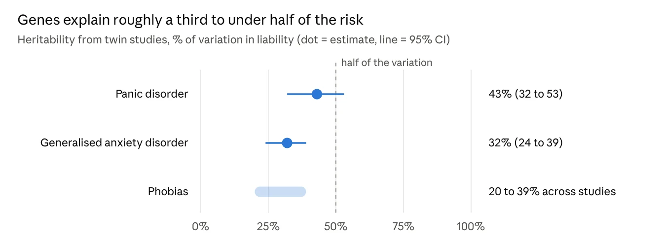
The part not explained by genes is mostly what twin researchers call non-shared environment: experiences that differ between members of the same family, for example a frightening event or an illness that only one sibling went through. The family environment that siblings share seemed to matter little for panic disorder. For generalised anxiety disorder a small shared-environment effect appeared in women only and the authors judged it uncertain.
Finding the genes has proved slow. A meta-analysis of genome-wide association studies across nine samples and more than 18,000 people found one genome-wide significant region for each way of defining the phenotype, but none of them had been replicated (Otowa et al., 2016). The likely explanation is that risk is spread across many variants, each with a tiny effect.
Epigenetics offers a way to link genes and experience. In rats, pups that receive more licking and grooming from their mother show lower DNA methylation at the promoter of the glucocorticoid receptor gene in the hippocampus. They express more receptors, shut down the stress response more efficiently and stay calmer as adults. Cross-fostering showed that the effect follows the mother's care, not the pup's genes (Weaver et al., 2004). A post-mortem study found a similar pattern in humans: in the hippocampus of suicide victims who had been abused as children, the same gene was more methylated and less expressed than in suicide victims without abuse or in controls (McGowan et al., 2009). Each group had only 12 people.
The clearest example of gene and environment acting together involves FKBP5, a gene that regulates sensitivity to cortisol. In carriers of a risk variant, childhood trauma, but not trauma in adulthood, was linked to a loss of methylation in a regulatory region of the gene and to lasting changes in the stress-hormone system (Klengel et al., 2013). This work concerned post-traumatic stress and depression more than the anxiety disorders discussed here and direct epigenetic evidence for panic or generalised anxiety is still thin. The principle, though, is general: early adversity can change how stress genes are read and the timing of the adversity matters.
Clinical implications
Neurobiology cannot yet diagnose an anxiety disorder or say which treatment suits a given person, but it gives patients a model they can understand. The alarm runs on old, fast circuits that are hard to switch off by willpower. The prefrontal brake can be trained and exposure therapy trains it by building a new safety memory next to the old fear memory. For a frightened patient, knowing that the old memory stays and can be outcompeted may make exposure easier to accept.
Several findings once presented as established have shrunk or become more complicated with larger samples. Two examples are the amygdala as the single fear centre and the serotonin transporter gene as a risk marker.
I see clients in English, French and Italian, at the practice in Gasperich (Luxembourg) on Saturdays or by video consultation on weekday evenings.
Request an appointmentReferences
- Avery, S. N., Clauss, J. A., & Blackford, J. U. (2016). The human BNST: Functional role in anxiety and addiction. Neuropsychopharmacology, 41(1), 126–141. https://doi.org/10.1038/npp.2015.185
- Baldwin, D. S., Anderson, I. M., Nutt, D. J., Allgulander, C., Bandelow, B., den Boer, J. A., Christmas, D. M., Davies, S., Fineberg, N., Lidbetter, N., Malizia, A., McCrone, P., Nabarro, D., O'Neill, C., Scott, J., van der Wee, N., & Wittchen, H.-U. (2014). Evidence-based pharmacological treatment of anxiety disorders, post-traumatic stress disorder and obsessive-compulsive disorder: A revision of the 2005 guidelines from the British Association for Psychopharmacology. Journal of Psychopharmacology, 28(5), 403–439. https://doi.org/10.1177/0269881114525674
- Bandelow, B., Baldwin, D., Abelli, M., Altamura, C., Dell'Osso, B., Domschke, K., Fineberg, N. A., Grünblatt, E., Jarema, M., Maron, E., Nutt, D., Pini, S., Vaghi, M. M., Wichniak, A., Zai, G., & Riederer, P. (2016). Biological markers for anxiety disorders, OCD and PTSD: A consensus statement. Part I: Neuroimaging and genetics. The World Journal of Biological Psychiatry, 17(5), 321–365. https://doi.org/10.1080/15622975.2016.1181783
- Binder, E. B., & Nemeroff, C. B. (2010). The CRF system, stress, depression and anxiety: Insights from human genetic studies. Molecular Psychiatry, 15(6), 574–588. https://doi.org/10.1038/mp.2009.141
- Davis, M., Walker, D. L., Miles, L., & Grillon, C. (2010). Phasic vs sustained fear in rats and humans: Role of the extended amygdala in fear vs anxiety. Neuropsychopharmacology, 35(1), 105–135. https://doi.org/10.1038/npp.2009.109
- Elnazer, H. Y., & Baldwin, D. S. (2014). Investigation of cortisol levels in patients with anxiety disorders: A structured review. Current Topics in Behavioral Neurosciences, 18, 191–216. https://doi.org/10.1007/7854_2014_299
- Etkin, A., Egner, T., & Kalisch, R. (2011). Emotional processing in anterior cingulate and medial prefrontal cortex. Trends in Cognitive Sciences, 15(2), 85–93. https://doi.org/10.1016/j.tics.2010.11.004
- Etkin, A., Prater, K. E., Hoeft, F., Menon, V., & Schatzberg, A. F. (2010). Failure of anterior cingulate activation and connectivity with the amygdala during implicit regulation of emotional processing in generalized anxiety disorder. American Journal of Psychiatry, 167(5), 545–554. https://doi.org/10.1176/appi.ajp.2009.09070931
- Etkin, A., Prater, K. E., Schatzberg, A. F., Menon, V., & Greicius, M. D. (2009). Disrupted amygdalar subregion functional connectivity and evidence of a compensatory network in generalized anxiety disorder. Archives of General Psychiatry, 66(12), 1361–1372. https://doi.org/10.1001/archgenpsychiatry.2009.104
- Etkin, A., & Wager, T. D. (2007). Functional neuroimaging of anxiety: A meta-analysis of emotional processing in PTSD, social anxiety disorder and specific phobia. American Journal of Psychiatry, 164(10), 1476–1488. https://doi.org/10.1176/appi.ajp.2007.07030504
- Fullana, M. A., Harrison, B. J., Soriano-Mas, C., Vervliet, B., Cardoner, N., Àvila-Parcet, A., & Radua, J. (2016). Neural signatures of human fear conditioning: An updated and extended meta-analysis of fMRI studies. Molecular Psychiatry, 21(4), 500–508. https://doi.org/10.1038/mp.2015.88
- Grupe, D. W., & Nitschke, J. B. (2013). Uncertainty and anticipation in anxiety: An integrated neurobiological and psychological perspective. Nature Reviews Neuroscience, 14(7), 488–501. https://doi.org/10.1038/nrn3524
- Hettema, J. M., Neale, M. C., & Kendler, K. S. (2001). A review and meta-analysis of the genetic epidemiology of anxiety disorders. American Journal of Psychiatry, 158(10), 1568–1578. https://doi.org/10.1176/appi.ajp.158.10.1568
- Klengel, T., Mehta, D., Anacker, C., Rex-Haffner, M., Pruessner, J. C., Pariante, C. M., Pace, T. W. W., Mercer, K. B., Mayberg, H. S., Bradley, B., Nemeroff, C. B., Holsboer, F., Heim, C. M., Ressler, K. J., Rein, T., & Binder, E. B. (2013). Allele-specific FKBP5 DNA demethylation mediates gene–childhood trauma interactions. Nature Neuroscience, 16(1), 33–41. https://doi.org/10.1038/nn.3275
- LeDoux, J. E., & Pine, D. S. (2016). Using neuroscience to help understand fear and anxiety: A two-system framework. American Journal of Psychiatry, 173(11), 1083–1093. https://doi.org/10.1176/appi.ajp.2016.16030353
- Malizia, A. L., Cunningham, V. J., Bell, C. J., Liddle, P. F., Jones, T., & Nutt, D. J. (1998). Decreased brain GABA(A)-benzodiazepine receptor binding in panic disorder: Preliminary results from a quantitative PET study. Archives of General Psychiatry, 55(8), 715–720. https://doi.org/10.1001/archpsyc.55.8.715
- McGowan, P. O., Sasaki, A., D'Alessio, A. C., Dymov, S., Labonté, B., Szyf, M., Turecki, G., & Meaney, M. J. (2009). Epigenetic regulation of the glucocorticoid receptor in human brain associates with childhood abuse. Nature Neuroscience, 12(3), 342–348. https://doi.org/10.1038/nn.2270
- Milad, M. R., & Quirk, G. J. (2012). Fear extinction as a model for translational neuroscience: Ten years of progress. Annual Review of Psychology, 63, 129–151. https://doi.org/10.1146/annurev.psych.121208.131631
- Munafò, M. R., Brown, S. M., & Hariri, A. R. (2008). Serotonin transporter (5-HTTLPR) genotype and amygdala activation: A meta-analysis. Biological Psychiatry, 63(9), 852–857. https://doi.org/10.1016/j.biopsych.2007.08.016
- Murphy, S. E., Norbury, R., Godlewska, B. R., Cowen, P. J., Mannie, Z. M., Harmer, C. J., & Munafò, M. R. (2013). The effect of the serotonin transporter polymorphism (5-HTTLPR) on amygdala function: A meta-analysis. Molecular Psychiatry, 18(4), 512–520. https://doi.org/10.1038/mp.2012.19
- Nuss, P. (2015). Anxiety disorders and GABA neurotransmission: A disturbance of modulation. Neuropsychiatric Disease and Treatment, 11, 165–175. https://doi.org/10.2147/NDT.S58841
- Otowa, T., Hek, K., Lee, M., Byrne, E. M., Mirza, S. S., Nivard, M. G., Bigdeli, T., Aggen, S. H., Adkins, D., Wolen, A., Fanous, A., Keller, M. C., Castelao, E., Kutalik, Z., Van der Auwera, S., Homuth, G., Nauck, M., Teumer, A., Milaneschi, Y., … Hettema, J. M. (2016). Meta-analysis of genome-wide association studies of anxiety disorders. Molecular Psychiatry, 21(10), 1391–1399. https://doi.org/10.1038/mp.2015.197
- Shackman, A. J., & Fox, A. S. (2016). Contributions of the central extended amygdala to fear and anxiety. Journal of Neuroscience, 36(31), 8050–8063. https://doi.org/10.1523/JNEUROSCI.0982-16.2016
- Tovote, P., Fadok, J. P., & Lüthi, A. (2015). Neuronal circuits for fear and anxiety. Nature Reviews Neuroscience, 16(6), 317–331. https://doi.org/10.1038/nrn3945
- Vreeburg, S. A., Zitman, F. G., van Pelt, J., DeRijk, R. H., Verhagen, J. C. M., van Dyck, R., Hoogendijk, W. J. G., Smit, J. H., & Penninx, B. W. J. H. (2010). Salivary cortisol levels in persons with and without different anxiety disorders. Psychosomatic Medicine, 72(4), 340–347. https://doi.org/10.1097/PSY.0b013e3181d2f0c8
- Weaver, I. C. G., Cervoni, N., Champagne, F. A., D'Alessio, A. C., Sharma, S., Seckl, J. R., Dymov, S., Szyf, M., & Meaney, M. J. (2004). Epigenetic programming by maternal behavior. Nature Neuroscience, 7(8), 847–854. https://doi.org/10.1038/nn1276
© 2018 Giampaolo Penzo. All rights reserved. No reproduction, translation or reuse, including for AI training, without the author's written permission. Short quotations are welcome with credit and a link to this page. Legal notice